Peyronies disease ultrasound is sometimes requested by patients who contact Moorgate Andrology because they have noticed a change in the shape of their erection.
A change in the shape of the erection can be very alarming. Not only that, when the angle of the erection is quite acute, it can make sexual intercourse difficult or even impossible.
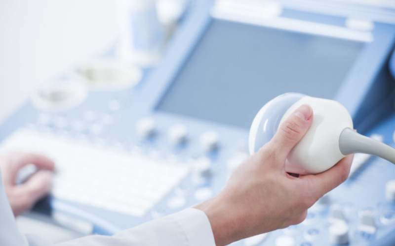
A trip to the Urologist’s office at Moorgate Andrology can indeed include an ultrasound scan of the penis. An ultrasound scan of the penis can help the Urologist highlight the areas of plaque that are the cause of the unnatural bending of the penis. He can also check for the blood flow as sometimes erections are also compromised to a greater or lesser extent.
An ultrasound scan of the penis is a painless experience. It takes between ten and twenty minutes.
Firstly, the Urologist will apply some gel to the penis and then he will apply the probe to the penis. He will move the probe around the shaft of the penis to get a detailed picture of the condition.
At this consultation, he may have asked you to bring along some photos of the penis in full erection to help with the overall assessment of the disease.
The ultrasound and photos will enable both you and your Urologist to decide the best course of action to get you back on the road to more normal erections.
Of course, the type of treatment, or surgery, will somewhat depend on the stage of your disease. Usually, surgical procedures are only undertaken when the disease has reached the chronic phase, which can take up to one year.
If you are in the acute phase then your Urologist may want to wait until the disease has reached the chronic phase before considering any surgical intervention. In the meantime, more conservative treatment may be considered to minimise the disease progression as much as possible.
Further periodic ultrasound scans can be undertaken to monitor your disease in the forthcoming months.


LEAVE A REPLY Cancel Reply
Your email address will not be published. Required fields are marked *